The Peripheral Nervous System—Nerves on the Edge
-
2.5 Describe the role of the somatic and autonomic nervous systems.
The term peripheral refers to things that are not in the center or that are on the edges of the center. The peripheral nervous system or PNS (see Figure 2.7 and also refer back to Figure 2.5) is made up of all the nerves and neurons that are not contained in the brain and spinal cord. It is this system that allows the brain and spinal cord to communicate with the sensory systems and enables the brain and spinal cord to control the muscles and glands of the body. The PNS can be divided into two major systems: the somatic nervous system, which consists of nerves that control the voluntary muscles of the body, and the autonomic nervous system (ANS), which consists of nerves that control the involuntary muscles, organs, and glands.
Figure 2.7
The Peripheral Nervous System

The Somatic Nervous System
One of the parts of a neuron is the soma, or cell body (remember that the word soma means “body”). The somatic nervous system is made up of the sensory pathway, which is all the nerves carrying messages from the senses to the central nervous system (those nerves containing afferent neurons), and the motor pathway, which is all the nerves carrying messages from the central nervous system to the voluntary, or skeletal,more info muscles of the body—muscles that allow people to move their bodies (those nerves composed of efferent neurons). When people are walking, raising their hands in class, smelling a flower, or directing their gaze toward the person they are talking to or to look at a pretty picture, they are using the somatic nervous system. (As seen in the discussion of spinal cord reflexes, although these muscles are called the voluntary muscles, they can move involuntarily when a reflex response occurs. They are called “voluntary” because they can be moved at will but are not limited to only that kind of movement.)
Involuntarymore info muscles, such as the heart, stomach, and intestines, together with glands such as the adrenal glands and the pancreas are all controlled by clumps of neurons located on or near the spinal column. (The words on or near are used quite deliberately here. The neurons inside the spinal column are part of the central nervous system, not the peripheral nervous system.) These large groups of neurons near the spinal column make up the autonomic nervous system.
The Autonomic Nervous System
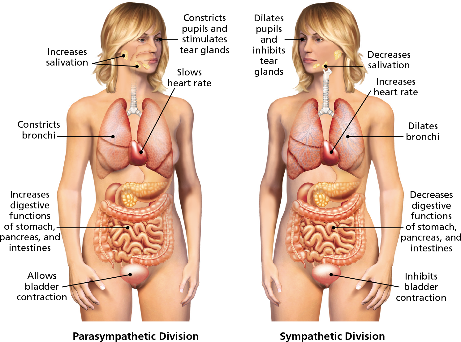
The word autonomic suggests that the functions of this system are more or less automatic, which is basically correct. Whereas the somatic division of the peripheral nervous system controls the senses and voluntary muscles, the autonomic division controls everything else in the body—organs, glands, and involuntary muscles. The autonomic nervous system is divided into two systems, the sympathetic division, and the parasympathetic division. (See Figure 2.8).
Figure 2.8
Functions of the Parasympathetic and Sympathetic Divisions of the Nervous System


These young soccer players are using their senses and voluntary muscles controlled by the somatic division of the peripheral nervous system. What part of the autonomic nervous system are these girls also using at this time?
The Sympathetic Division
The sympathetic division is usually called the “fight-or-flight system” because it allows people and animals to deal with all kinds of stressful events. (See Learning Objective 2.7.) Emotions during these events might be anger (hence, the term fight) or fear (that’s the “flight” part, obviously) or even extreme joy or excitement. Yes, even joy can be stressful. The sympathetic division’s job is to get the body ready to deal with the stress. Many of us have experienced a fight-or-flight moment at least once in our lives. We will look more closely at how the body responds to stress later in the chapter but for now, participate in the survey Do You Fly or Fight? to learn more about how your body responds in certain situations.
Survey
Do You Fly or Fight?
What are the specific ways in which this division readies the body to react? (See Figure 2.8.) The pupils seem to get bigger, perhaps to let in more light and, therefore, more information. The heart starts pumping faster and harder, drawing blood away from nonessential organs such as the skin (so at first the person may turn pale) and sometimes even the brain itself (so the person might actually faint). Blood needs lots of oxygen before it goes to the muscles, so the lungs work overtime, too (so the person may begin to breathe faster). One set of glands in particular receives special instructions. The adrenal glands will be stimulated to release certain stress-related chemicals (members of a class of chemicals released by glands called hormones) into the bloodstream. These stress hormones will travel to all parts of the body, but they will only affect certain target organs. Just as a neurotransmitter fits into a receptor site on a cell, the molecules of the stress hormones fit into receptor sites at the various target organs—notably, the heart, muscles, and lungs. This further stimulates these organs to work harder. But not every organ or system will be stimulated by the activation of the sympathetic division. Digestion of food and excretionmore info of waste are not necessary functions when dealing with stressful situations, so these systems tend to be “shut down” or inhibited. Saliva, which is part of digestion, dries right up (ever try whistling when you’re scared?). Food that was in the stomach sits there like a lump. Usually, the urge to go to the bathroom will be suppressed, but if the person is really scared the bladder or bowels may actually empty (this is why people who die under extreme stress, such as hanging or electrocution, will release their urine and waste). The sympathetic division is also going to demand that the body burn a tremendous amount of fuel, or blood sugar.
Now, all this bodily arousal is going on during a stressful situation. If the stress ends, the activity of the sympathetic division will be replaced by the activation of the parasympathetic division. If the stress goes on too long or is too intense, the person might actually collapse (as a deer might do when being chased by another animal). This collapse occurs because the parasympathetic division overresponds in its inhibition of the sympathetic activity. The heart slows, blood vessels open up, blood pressure in the brain drops, and fainting can result.
The Parasympathetic Division
If the sympathetic division can be called the fight-or-flight system, the parasympathetic division might be called the “eat-drink-and-rest” system. The neurons of this division are located at the top and bottom of the spinal column on either side of the sympathetic division neurons (para means “beyond” or “next to” and in this sense refers to the neurons located on either side of the sympathetic division neurons).
In looking at Figure 2.8, it might seem as if the parasympathetic division does pretty much the opposite of the sympathetic division, but it’s a little more complex than that. The parasympathetic division’s job is to restore the body to normal functioning after a stressful situation ends. It slows the heart and breathing, constricts the pupils, and reactivates digestion and excretion. Signals to the adrenal glands stop because the parasympathetic division isn’t connected to the adrenal glands. In a sense, the parasympathetic division allows the body to put back all the energy it burned—which is why people are often very hungry after the stress is all over.
The parasympathetic division does more than just react to the activity of the sympathetic division. It is the parasympathetic division that is responsible for most of the ordinary, day-to-day bodily functioning, such as regular heartbeat and normal breathing and digestion. People spend the greater part of their 24-hour day eating, sleeping, digesting, and excreting. So it is the parasympathetic division that is normally active. At any given moment, then, one or the other of these divisions, sympathetic or parasympathetic, will determine whether people are aroused or relaxed.
Concept Map 2.4
Click or Tap the Start Button to Explore the Concept and + to Expand.
Concept Map 2.5
Click or Tap the Start Button to Explore the Concept and + to Expand.