The Cortex
-
2.12 Identify the parts of the cortex that process the different senses and those that control movement of the body.
As stated earlier, the cortex (“rind” or outer covering) is the outermost part of the brain, which is the part of the brain most people picture when they think of what the brain looks like. It is made up of tightly packed neurons and actually is only about one-tenth of an inch thick on average (Fischl et al., 2001; MacDonald et al., 2000; Zilles, 1990). The cortex has a very recognizable surface anatomy because it is full of wrinkles.
Why is the cortex so wrinkled?
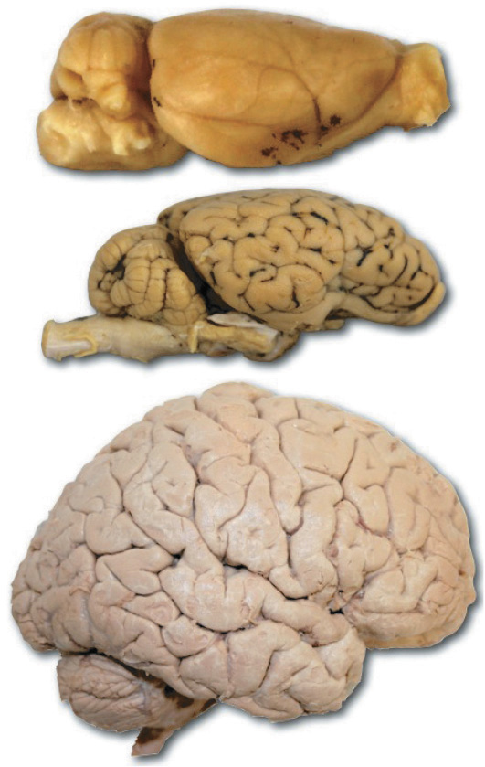
The wrinkling of the cortex allows a much larger area of cortical cells to exist in the small space inside the skull. If the cortex were to be taken out, ironed flat, and measured, it would be about 2 to 3 square feet. (The owner of the cortex would also be dead, but that’s fairly obvious, right?) As the brain develops before birth, it forms a smooth outer covering on all the other brain structures. This will be the cortex, which will get more and more wrinkled as the brain increases in size and complexity. This increase in wrinkling is called “corticalization.”

From top to bottom, a rat brain, sheep brain, and human brain (not to scale!). Note the differences in the amount of corticalization, or wrinkling, of the cortex between these three brains. Greater amounts of corticalization are associated with increases in size and complexity.
Cerebral Hemispheres
The brain is divided into two sections called the cerebral hemispheres, which are connected by a thick, tough band of neural fibers (axons) called the corpus callosum (literally meaning “hard bodies,” as calluses on the feet are hard). (Refer back to Figure 2.15.) The corpus callosum allows the left and right hemispheres to communicate with each other. Each hemisphere can be roughly divided into four sections or lobes by looking at the deeper wrinkles, or fissures, in its surface. The lobes are named for the skull bones that cover them (see Figure 2.16).
Figure 2.16
The Lobes of the Brain: Frontal, Temporal, Parietal, and Occipital
Another organizational feature of the cortex is that for specific regions, each hemisphere is responsible for the opposite side of the body, either for control, or for receiving information. For example, the motor cortex controls the muscles on the opposite side of the body. If we are writing with our right hand, the motor cortex in the left hemisphere is responsible for controlling those movements. This feature, referred to as contralateral organization, plays a role in information coming from many of the sense organs to the brain, and in the motor commands originating in the brain going to the rest of the body.
Information from our body can also be transmitted to both sides of the brain, or bilaterally (as in hearing and vision), or to only one side of the brain, or ipsilaterally (as in taste and olfaction). These aspects are also important in the study of brain lateralization, which we will come back to later in the chapter. Why do we have this arrangement for some functions and not for others? No one really knows, but at least for some information, it assists with identifying where information from the environment is coming from. For auditory information from the ears, having sensory information projected to both hemispheres allows us to localize sounds by comparing the slightly different information coming from each ear.
Occipital Lobes
At the base of the cortex, toward the back of the brain is an area called the occipital lobe. This area processes visual information from the eyes in the primary visual cortex. The visual association cortex, also in this lobe, is the part of the brain that helps identify and make sense of the visual information from the eyes. The famed neurologist Oliver Sacks once had a patient who had a tumor in his right occipital lobe area. He could still see objects perfectly well and even describe them in physical terms, but he could not identify them by sight alone. For example, Sacks once gave him a rose to look at. The man turned it around and around and began to describe it as a “red inflorescence” of some type with a green tubular projection. Only when he held it under his nose (stimulating the sense of smell) did he recognize it as a rose (Sacks, 1990). Each area of the cortex has these association areas that help people make sense of sensory information.
Parietal Lobes
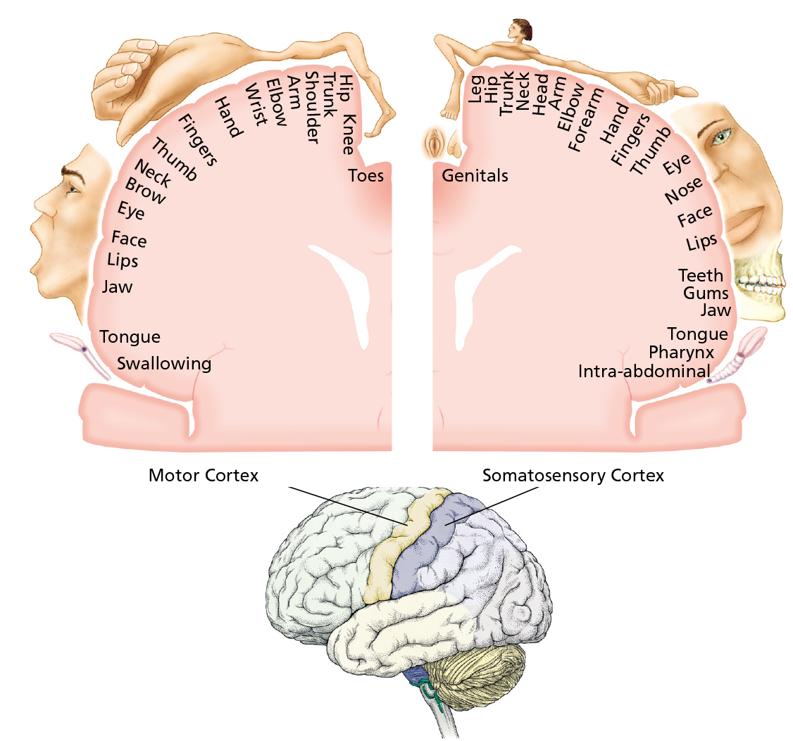
The parietal lobes are at the top and back of the brain, just under the parietal bone in the skull. This area contains the somatosensory cortex, an area of neurons (see Figure 2.17) at the front of the parietal lobes on either side of the brain. This area processes information from the skin and internal body receptors for touch, temperature, and body position. The somatosensory cortex is laid out in a rather interesting way—the cells at the top of the brain receive information from the bottom of the body, and as one moves down the area, the signals come from higher and higher in the body. It’s almost as if a little upside-down person were laid out along this area of cells.
Figure 2.17
The Motor and Somatosensory Cortex

The motor cortex in the frontal lobe controls the voluntary muscles of the body. Cells at the top of the motor cortex control muscles at the bottom of the body, whereas cells at the bottom of the motor cortex control muscles at the top of the body. Body parts are drawn larger or smaller according to the number of cortical cells devoted to that body part. For example, the hand has many small muscles and requires a larger area of cortical cells to control it. The somatosensory cortex, located in the parietal lobe just behind the motor cortex, is organized in much the same manner and receives information about the sense of touch and body position.
Temporal Lobes
The beginning of the temporal lobes are found just behind the temples of the head. These lobes contain the primary auditory cortex and the auditory association area. Also found in the left temporal lobe is an area that in most people is particularly involved with language. We have already discussed some of the medial structures of the temporal lobe, the amygdala and hippocampus, that are involved in aspects of learning and memory. There are also parts of the temporal lobe that help us process visual information.
Frontal Lobes
These lobes are at the front of the brain, hence, the name frontal lobes. (It doesn’t often get this easy in psychology; feel free to take a moment to appreciate it.) Here are found all the higher mental functions of the brain—planning, personality, memory storage, complex decision making, and (again in the left hemisphere in most people) areas devoted to language. The frontal lobe also helps in controlling emotions by means of its connection to the limbic system. The most forward part of the frontal lobes is called the prefrontal cortex. The middle area toward the center (medial prefrontal cortex) and bottom surface above the eyes (orbitofrontal prefrontal cortex—right above the orbits of the eye) have strong connections to the limbic system. Phineas Gage, who was mentioned in Chapter One, suffered damage to his left frontal lobe. He lacked emotional control because of the damage to his prefrontal cortex and the connections with limbic system structures. Overall, he had connections damaged from the left frontal cortex to many other parts of the brain (Ratiu et al., 2004; Van Horn et al., 2012). People with damage to the frontal lobe may also experience problems with performing mental or motor tasks, such as getting stuck on one step in a process or on one wrong answer in a test and repeating it over and over again, or making the same movement over and over, a phenomena called perseveration (Asp & Tranel, 2013; Luria, 1965; Goel & Grafman, 1995).

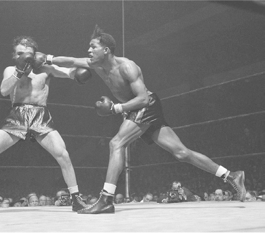
This boxer must rely on his parietal lobes to sense where his body is in relation to the floor of the ring and the other boxer, his occipital lobes to see his target, and his frontal lobes to guide his hand and arm into the punch.
The frontal lobes also contain the motor cortex, a band of neurons located at the back of each lobe. (See Figure 2.17.) These cells control the movements of the body’s voluntary muscles by sending commands out to the somatic division of the peripheral nervous system. The motor cortex is laid out just like the somatosensory cortex, which is right next door in the parietal lobes.
This area of the brain has been the focus of a great deal of recent research, specifically as related to the role of a special type of neuron. These neurons are called mirror neurons, which fire when an animal performs an action—but they also fire when an animal observes that same action being performed by another. Previous brain-imaging studies in humans suggested that we, too, have mirror neurons in this area of the brain (Buccino et al., 2001; Buccino et al., 2004; Iacoboni et al., 1999). However, recent single-cell and multicell recordings in humans have demonstrated that neurons with mirroring functions are found not only in motor regions but also in parts of the brain involved in vision and memory. This suggests that such neurons provide much more information than previously thought about our own actions as compared to the actions of others (Mukamel et al., 2010). These findings may have particular relevance for better understanding or treating specific clinical conditions that are believed to involve a faulty mirror system in the brain such as autism (Oberman & Ramachandran, 2007; Rizzolatti et al., 2009). (See Learning Objective 8.8.)

As this boy imitates the motions his father goes through while shaving, certain areas of his brain are more active than others, areas that control the motions of shaving. But even if the boy were only watching his father, those same neural areas would be active—the neurons in the boy’s brain would mirror the actions of the father he is observing.
You’ve mentioned association cortex a few times. Do the other lobes of the brain contain association cortex as well?